This report concerns two cases of tuberculous pleural effusion in which significant numbers of mesothelial cells were found.
Mesothelial cells in pleural fluid tb or not tb.
The cytology showed reactive mesothelial cells and the differential cell count was as follows.
Tb or not tb.
Shapiro summary eighty five samples of pleural fluid obtained from 76.
Tb or not tb.
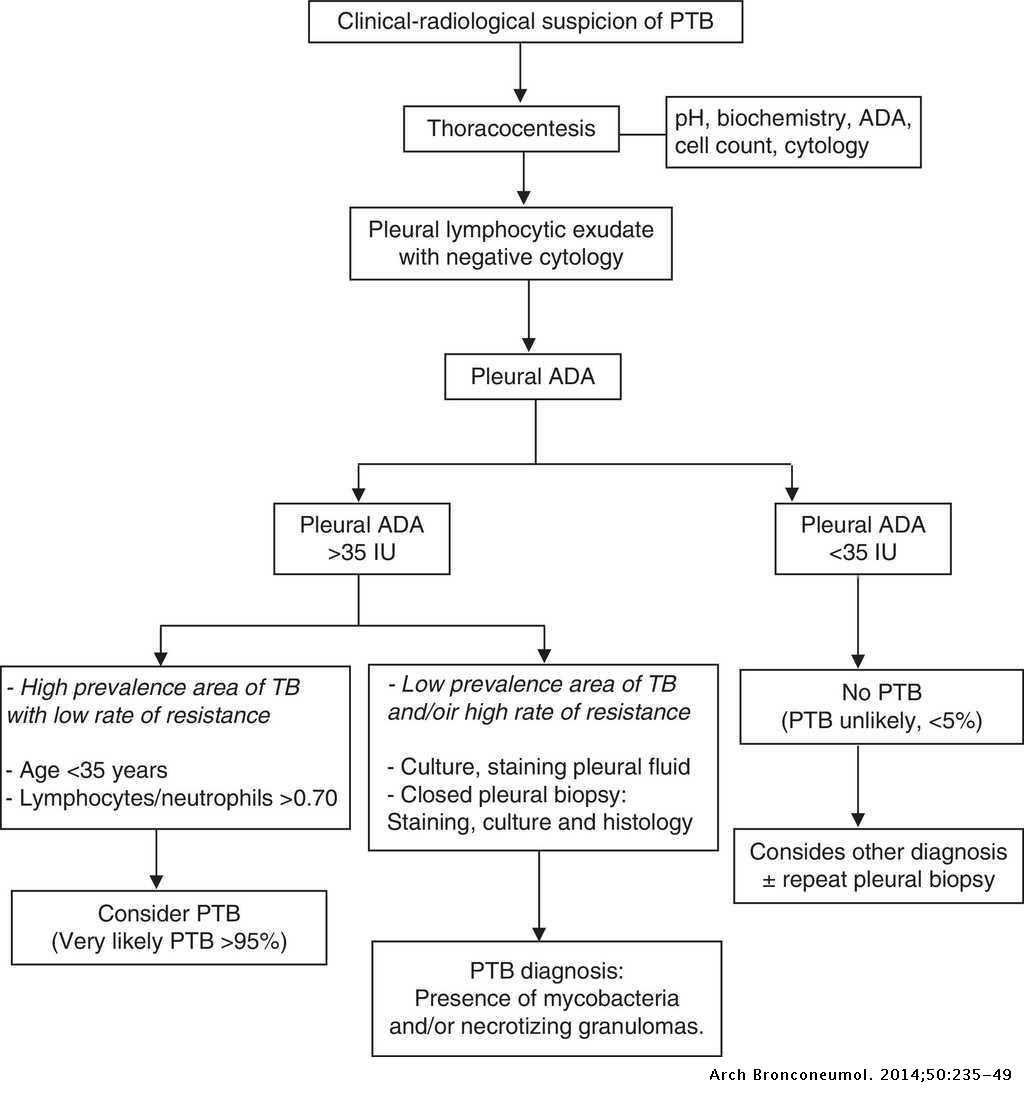
The diagnosis can be established in a majority of patients from the clinical features pleural fluid examination including cytology biochemistry and bacteriol ogy and pleural biopsy.
Use of pleural fluid n.
During development the mesoderm maintains a complex relationship with the developing endoderm giving rise to the mature lung.
Oa south african medical journal mesothelial cells in pleural fluid.
Navigate this journal about current issue previous issues submit a paper contact the editor oa african journal archive issn.
Mesothelial cells are found in variable numbers in most effusions but their presence at greater than 5 of total nucleated cells makes a diagnosis of tb less likely.
The culture of the pleural fluid grew m tuberculosis.
The patient was treated with a combination of six anti tb medications and was started on an antiretroviral regimen.
In contrast 65 3 of pleural fluid aspirates obtained from a control group of patients in congestive cardiac failure contained marked mesothelial exfoliation.
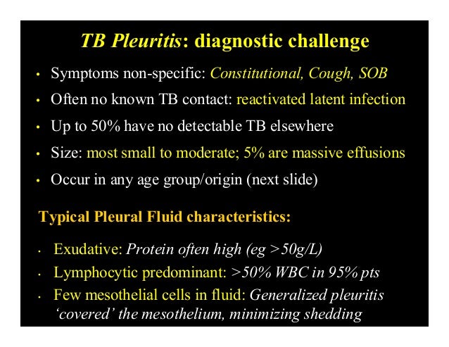
1 the fluid is generally an exudate 2 characterized by a predominance of lymphocytes and a paucity or absence of mesothelial cells 3 4 5 in fact it has been concluded that the presence of numerous mesothelial cells almost excludes a diagnosis of tuberculosis.
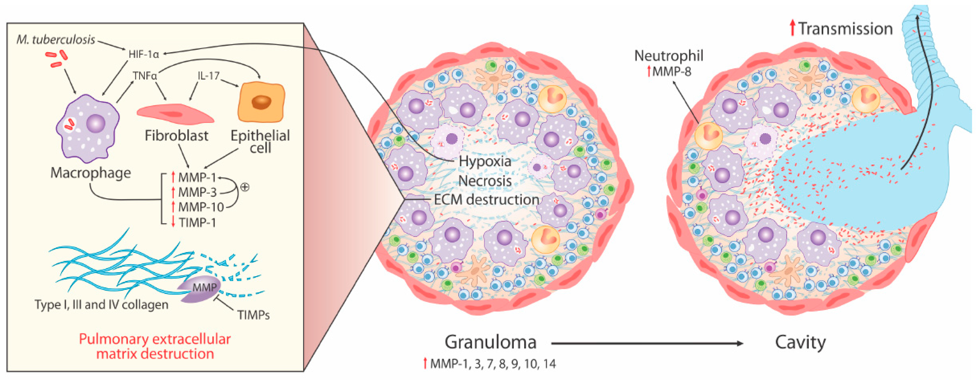
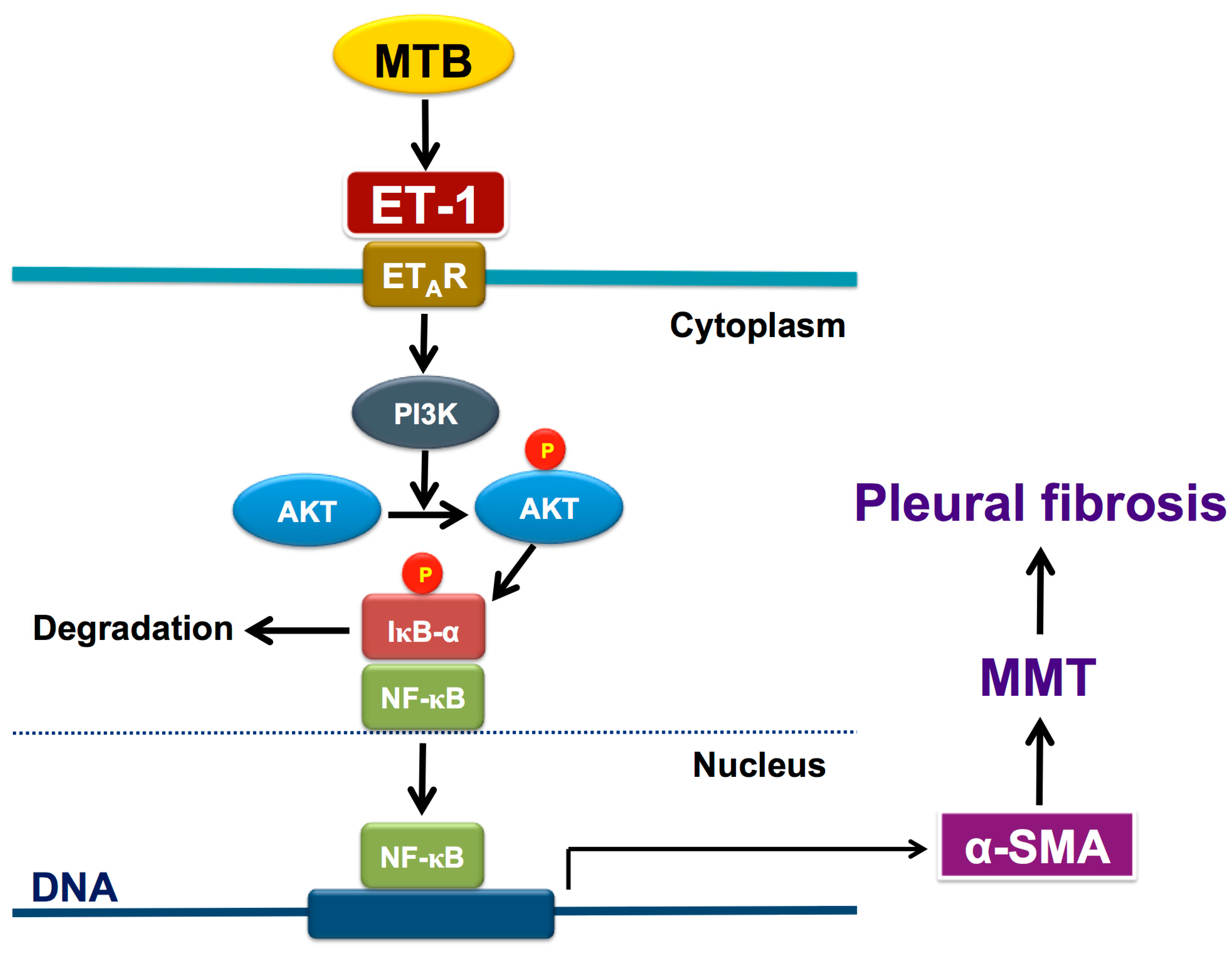
The pleural mesothelium differentiates to give rise to the endothelium and smooth muscle cells via epithelial to mesenchymal transition emt.
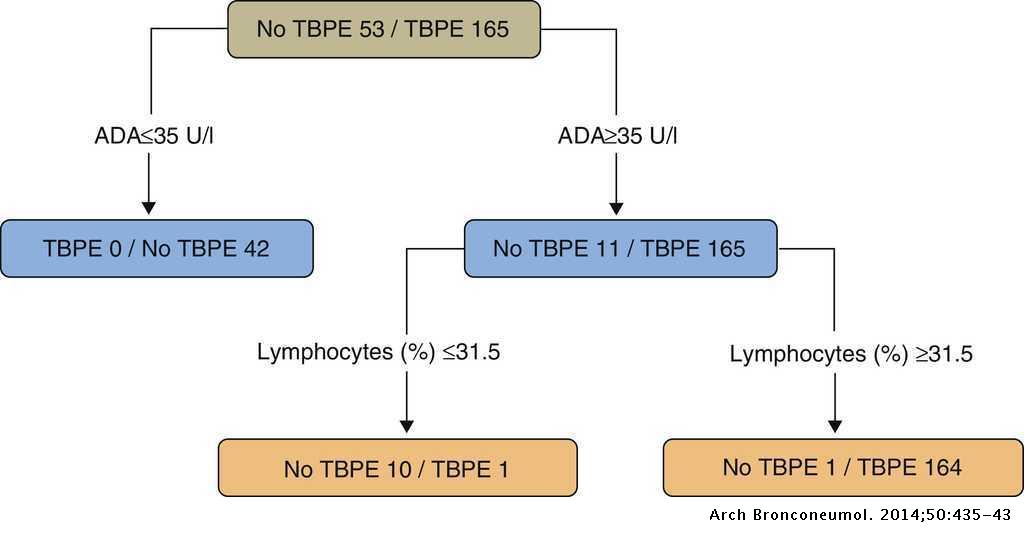
The presence of numerous mesothelial cells or eosinophils in the pleural fluid samples was useful to exclude tuberculosis in the differential diagnosis of exudative pleural effusions.
Based on the data of this study there is the possibility that mesothelial cells change to fibroblast like cells through emt in the process of tuberculous pleurisy.
7 june 1980 sa medical journal 937 m esothelial cells in pleural fluid.
Pleural mesothelial cells pmcs derived from the mesoderm play a key role during the development of the lung.
7 neutrophils 22 lymphocytes 60 macrophages and 10 mesothelial cells.
Therefore our finding could suggest that mesothelial cells undergo an epithleial to mesenchymal transition in the course of tuberculous pleurisy.
Pleural effusion may occur at any stage of active tuberculosis.
Although it has been accepted that effluents from tuberculous pleurisy rarely contain more than 5 mesothelial cells 35 36 the reasons have not yet been determined.


